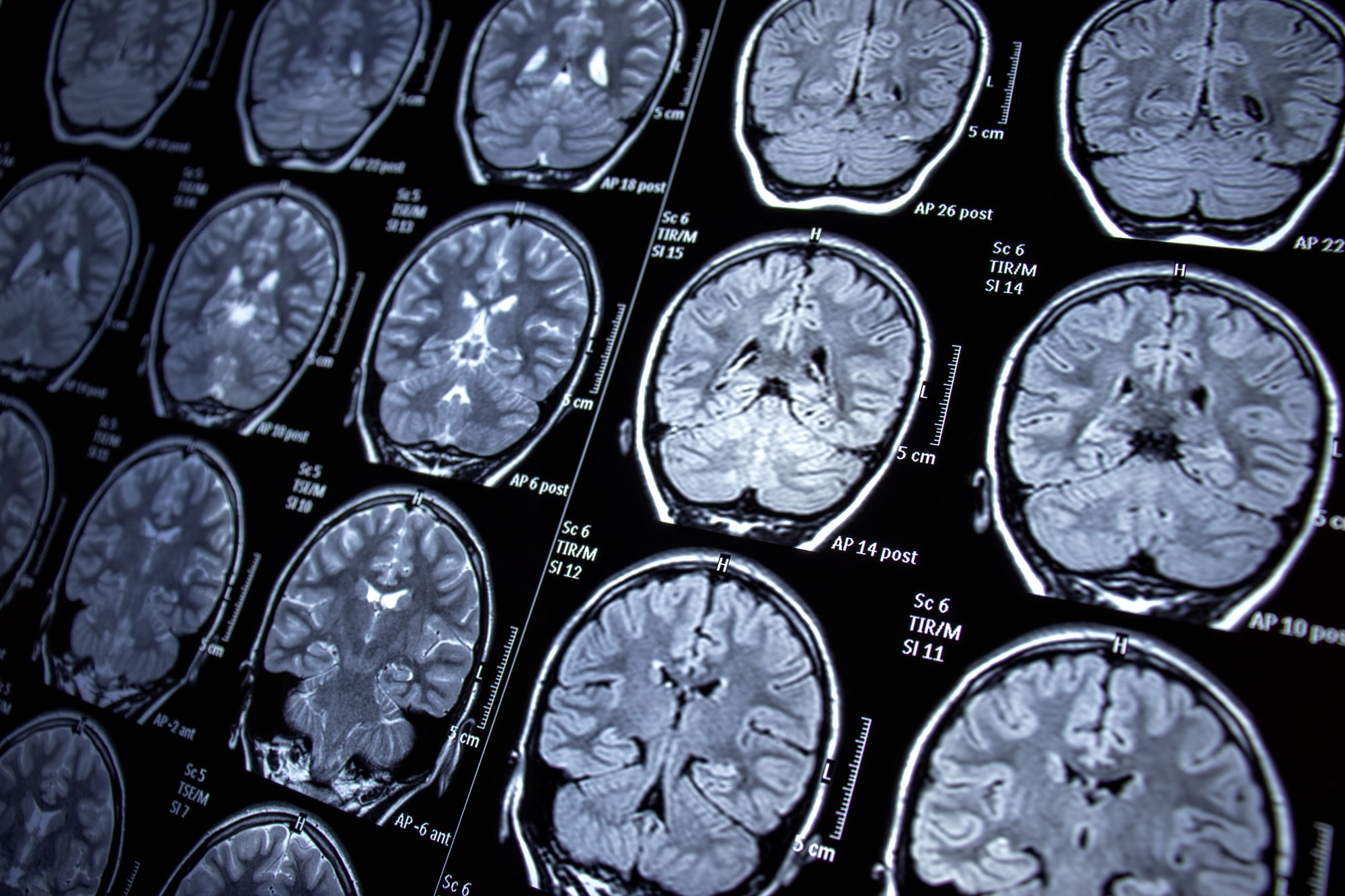
A new brain scanning technique developed at UBC can show how concussions damage the brain. These findings question the effectiveness of the tools doctors currently use when assessing and treating concussions.
To test this technique, researchers scanned the brains of male and female UBC hockey players before the start of the season. Multiple scans were done on 11 players who suffered a concussion. This study analyzed magnetic resonance imaging (MRI) to detect damage to the myelin sheath of hockey players.
Myelin is an insulating layer or sheath, that forms around the nerve fibre or axon in the brain. Myelin allows electrical impulses to transmit quickly and efficiently along the nerve cells. If myelin is damaged these impulses slow down.
Lead investigator Alex Rauscher found that the myelin is still damaged at two weeks. So even if the athlete passes the neuropsychological tests, he would keep him out of play for at least three weeks. He stated that “getting another concussion before the brain is healed can be extremely dangerous.”
The findings were published in the journals PLOS One and Frontiers in Neurology. A separate study published in the Canadian Medical Association Journal showed that only 31% of NHL players took more than 10 days off following a concussion.
Hopefully, Rauscher’s research encourages the leagues to modify their concussion protocols to protect their players.
For More Information
- UBC Researcher’s Scanning Technique Sheds New Light on Damage Done to the Brain by Concussion, The Province
- New Scanning Technique Reveals How Concussion Damages the Brain, The Vancouver Sun
- UBC News, UBC


