Imagine you’re Sydney Crosby. Or David Booth. Or Daniel Sedin. You are checked into the boards and your head hits the ice. Dazed, you are carried off the ice. Over the next few days, you suffer dizzy spells and headaches. These symptoms lead the team doctor to diagnose a concussion. But the doctor can’t tell you when you will recover or what limitations you may have. You just have to wait it out.
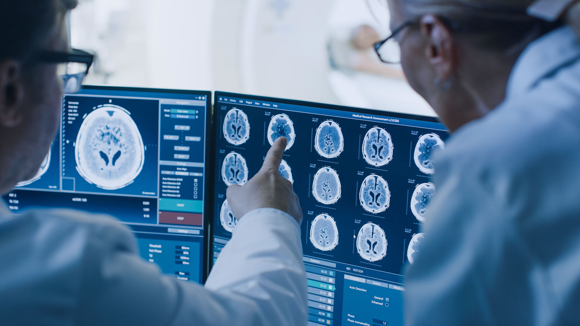
So why do some professional hockey players return to play within a few days while others have to retire from the game? In the area of brain injuries, current CT and MRI scans are not powerful enough to show detailed brain damage, making treatment and recovery much more difficult.
Dr. David Okonkwo, a neurosurgeon from the University of Pittsburgh Medical Center explains, “You can have a patient with severe swelling who goes on to have a normal recovery, and patients with severe swelling who go on to die.”
An article from the Associated Press reports that Dr. Okonkwo and a team of researchers are testing a new kind of MRI-based brain scan that is able to light up breaks in the brain’s axon connections. Known as high-definition fibre tracking (HDFT), the scan enables doctors to map a patient’s brain to determine possible long-term head injury effects. “It’s like comparing your fuzzy screen black-and-white TV with a high-definition TV,” says neurosurgeon Dr. Rocco Armonda.
So what are the advantages of seeing brain damage in HD? Like an HD TV, the HDFT shows details that highlight the damage unseen by other scans.
Take 32-year-old Daniel Stunkard, who spent three weeks in a coma after a devastating ATV accident. While CT and regular MRI scans showed some bruising and swelling to Stunkard’s brain, they were unable to indicate whether or not he would survive or what the extent of his injuries would be.
When Stunkard did wake up from his coma, specialists credit the new HDFT scan with predicting how his recovery would unfold. The scan revealed partial breaks in the nerve fibres controlling his leg and arm, but more extensive damage to the nerve fibres controlling his hand. Ten months later Stunkard is walking and has movement in his arm, but can’t use his left hand due to the more extensive nerve fibre damage shown on the scan.
This “game-changing” technology will improve concussion diagnosis in the years to come. “We now have, for the first time, the ability to make visible these previously invisible wounds,” says Walter Schneider, the researcher developing the scan. “If you cannot see or quantify the damage, it is hard to treat it.”
For More Information:
- Researchers aim to find invisible damage traumatic brain injury leaves behind, The Associated Press
- Concussion Questions and Answers, ThinkFirst Canada
- New Brain Imaging Technique Reveals Damage Caused by TBI, The University of Pittsburgh School of Health Science
- New High Definition Fiber Tracking Reveals Damage Caused by Traumatic Brain Injury, Science Daily


